投审稿入口


Research on CT image segmentation and identification methods for micro/nanoscale pores and fractures in sandstone
-
摘要:
致密砂岩微−纳米孔裂隙的精准识别是揭示岩石内部多相介质作用机制的重要基础。传统图像分割方法处理复杂孔裂隙结构时分割精度存在明显短板,且不同分割方法对孔隙三维重建、渗流分析结果的影响尚缺少系统评估。采用微焦点 X 射线计算机断层扫描(μCT)技术,构建致密砂岩微−纳米孔裂隙 CT 数据集;以灰度阈值分割、分水岭算法作为传统对照,选取 U-Net、SegNet、DeepLabv3-ResNet50、DeepLabv3-ResNet101共4种深度学习模型开展孔裂隙分割,同时引入公开微米砂岩数据集开展跨尺度迁移测试,综合对比不同方法二维分割精度、三维孔隙重建效果与渗流模拟结果。深度学习方法整体优于传统分割算法,其中 U-Net 模型综合性能最优,相较于传统方法最优结果,交并比(IoU)与F1分数分别提升 18.70%,16.47%,准确率达 99.03%,表现出较好的孔裂隙识别能力;U-Net 可较好保留纳米孔裂隙边界完整性与细节连续性,三维重建孔隙率为 2.408%,与原始孔隙率 2.785% 接近,重建的孔隙网络模型(PNM)孔喉参数与真实样本匹配度高。数据增强会破坏裂隙原生拓扑,造成模型精度下降;DeepLabv3 系列存在过分割问题,传统方法易出现微小孔隙漏检或虚假连通,进一步造成渗透率与压力场模拟偏差。相较于传统分割方法,U-Net 分割得到的孔隙网络拓扑与真实结构高度吻合,能够显著提升孔隙率、孔喉及渗透特性的定量分析精度,可为致密砂岩微−纳米孔裂隙自动识别与建模提供参考依据。
Abstract:ObjectiveAccurate characterization of micro/nanoscale pores and fractures in tight sandstone is fundamental for revealing the mechanisms of multiphase interactions within rocks, and it provides support for hydrocarbon exploitation, geothermal development, and geohazard assessment. Nevertheless, conventional image segmentation techniques suffer from obvious accuracy limitations when handling intricate pore-fracture structures. Moreover, few studies have systematically evaluated how segmentation algorithms affect subsequent three-dimensional pore reconstruction and seepage simulation results.
MethodsIn this study, micro-focus X-ray computed tomography (μCT) was adopted to build a self-established CT dataset containing micro/nanoscale pores and fractures of tight sandstone. Two conventional segmentation approaches—grayscale thresholding and the watershed algorithm—were employed as baseline comparisons. Four convolutional-neural-network-based deep learning models including U-Net, SegNet, DeepLabv3-ResNet50, and DeepLabv3-ResNet101 were implemented for pore-fracture segmentation. A publicly available micrometer-scale sandstone dataset was further utilized to conduct cross-scale transfer validation. Model performance was comprehensively compared and evaluated in terms of two-dimensional segmentation accuracy, three-dimensional pore network model (PNM) reconstruction, and single-phase seepage simulation.
ResultsThe experimental results showed that deep learning models outperformed traditional segmentation methods in general. Among all tested models, the U-Net achieved optimal comprehensive performance. Compared with the best results obtained using conventional segmentation methods, U-Net increased the intersection over union (IoU) and F1-score by 18.70% and 16.47%, respectively, with an overall accuracy of 99.03%, demonstrating favorable capability for pore and fracture identification. U-Net could effectively preserve the boundary integrity and fine-scale continuity of nanoscale pores and fractures. Its three-dimensional reconstructed porosity reached 2.408%, which was close to the original porosity of 2.785%, and the pore-throat parameters of the reconstructed pore network model (PNM) exhibited good consistency with the actual sample. It was noteworthy that data augmentation operations could destroy the inherent topological features of natural fractures and reduce model segmentation accuracy. The DeepLabv3 series models suffered from obvious over-segmentation artifacts, while traditional algorithms tended to produce missed detection of tiny pores or spurious pore connectivity. These defects further induced non-negligible deviations in permeability calculation and pressure-field distribution during seepage simulation.
ConclusionCompared with traditional segmentation approaches, the pore-network topology derived from U-Net segmentation matches well with real pore-fracture structures. The U-Net model substantially improves the quantitative analysis accuracy of porosity, pore-throat geometry, and permeability properties. This study provides a feasible reference for automatic identification and digital modeling of micro/nanoscale pore-fracture systems within tight sandstone.
-
Key words:
- pore-fracture segmentation /
- deep learning /
- CT images /
- tight sandstone /
- micro/nanoscale
-
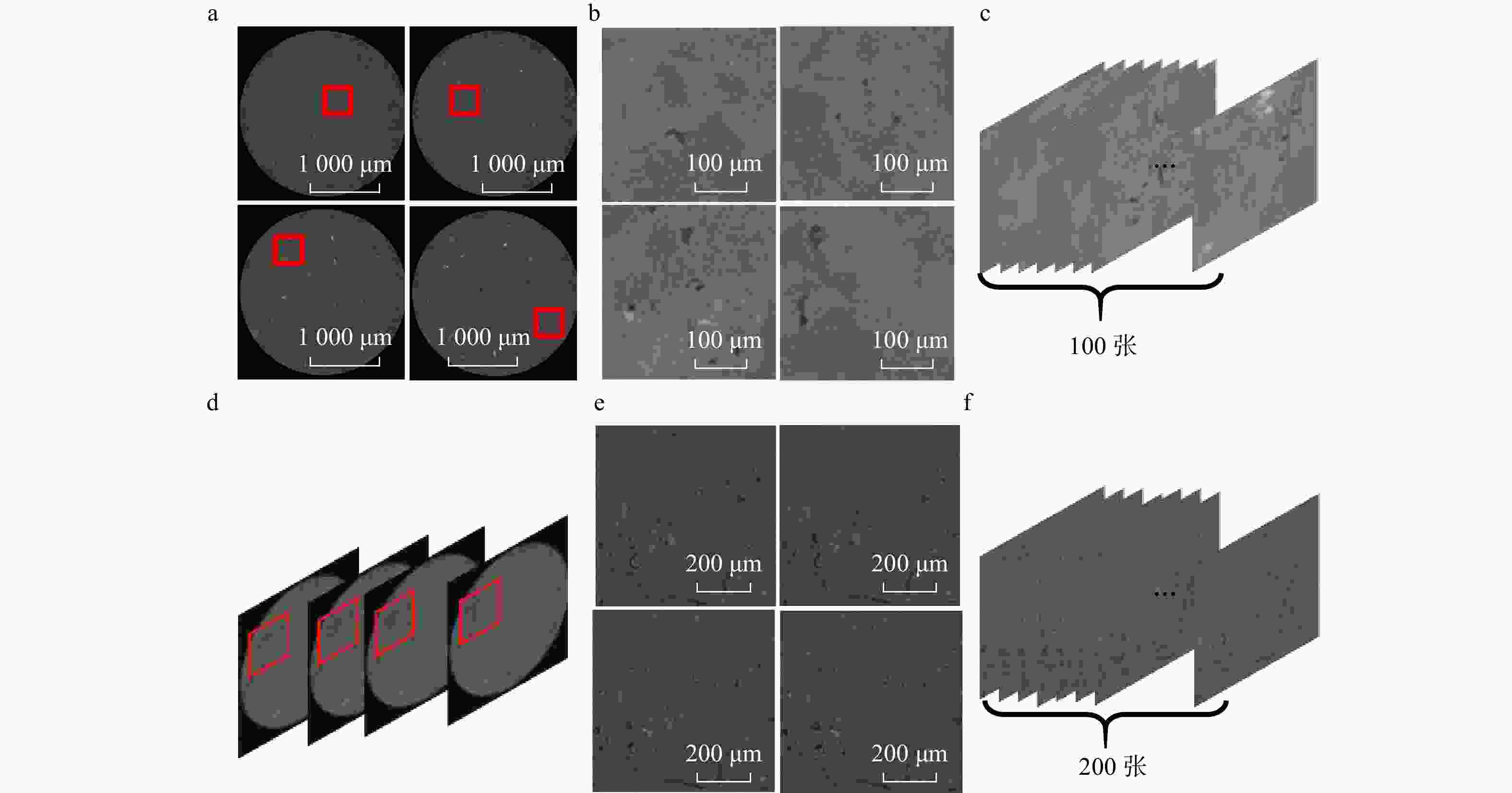
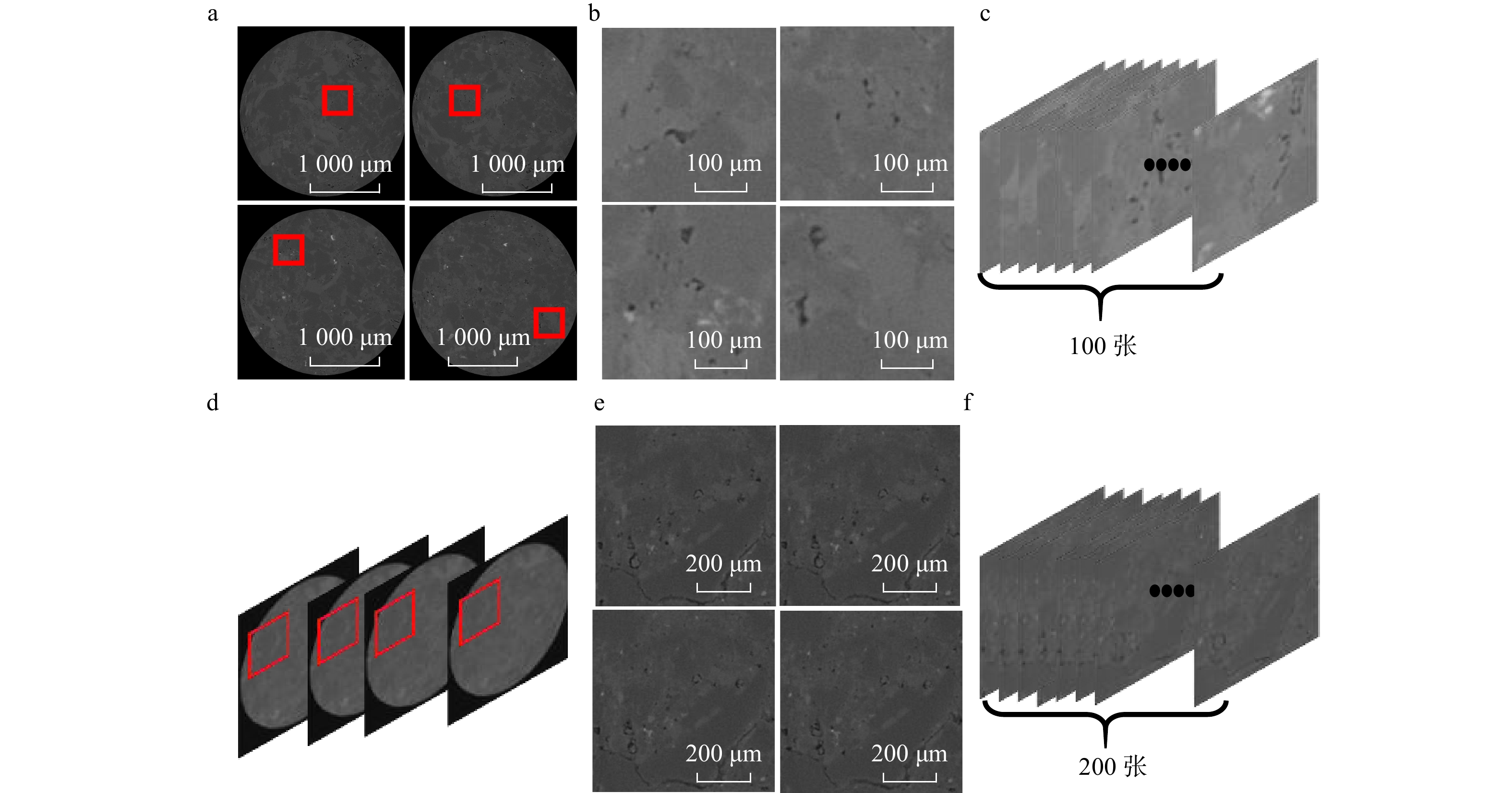
图 1 样本区域选取示意图
a为数据集一原始CT图像(红框标记待截取区域);b为截取得到的150 × 150像素、含显著裂隙的图像区域;c为数据集一,共100张图像;d为连续 CT 切片(红框标注同一截取位置);e为从连续切片同一位置截取的 300×300 像素图像区域;f为数据集二,共 200 张图像
Figure 1. Schematic diagram of sample area selection
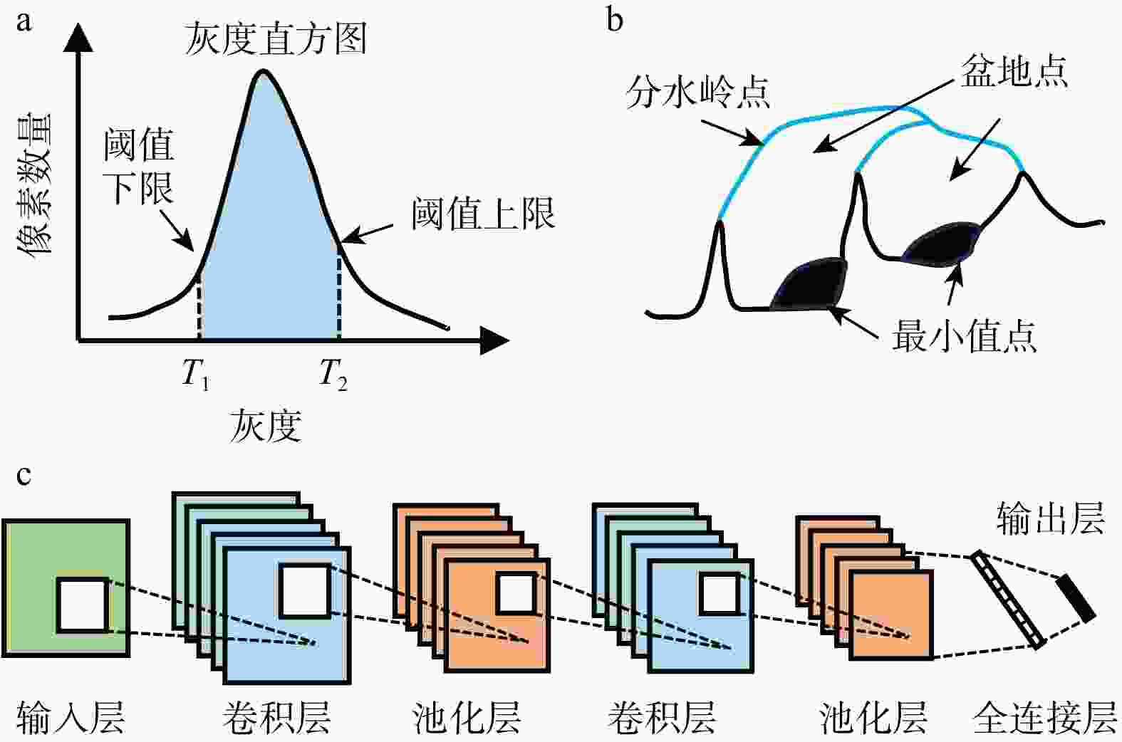
图 4 分割原理示意图(灰度区间T1~T2代表孔隙区域)
a. 基于图像灰度的交互式阈值(灰度阈值)分割;b. 分水岭概念;c. 卷积神经网络结构
Figure 4. Schematic diagram of segmentation principles
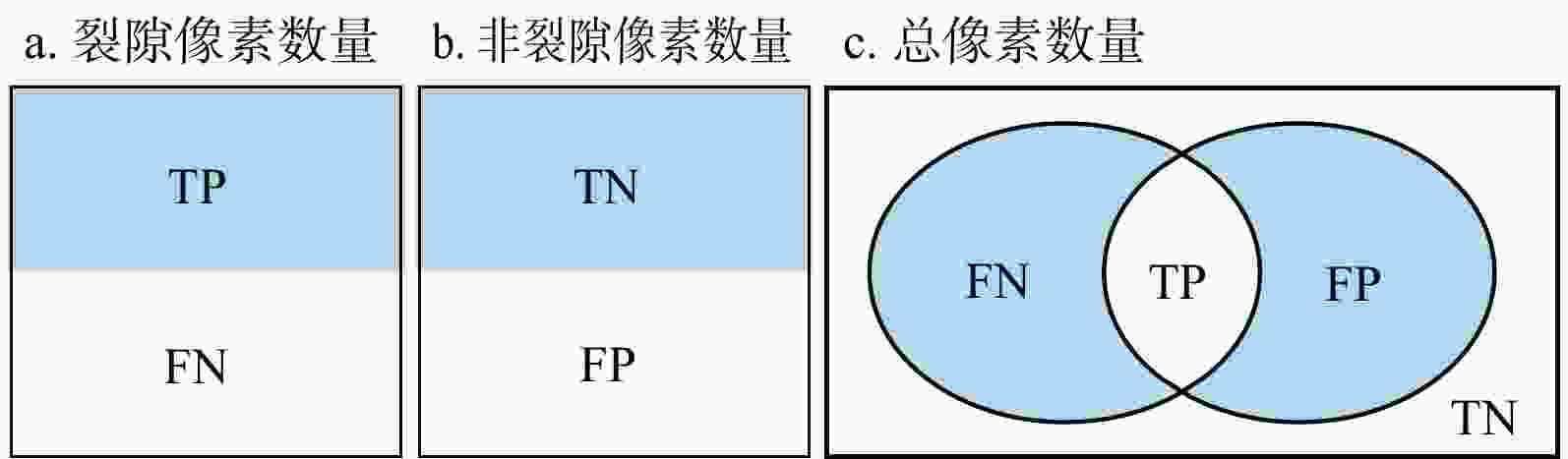
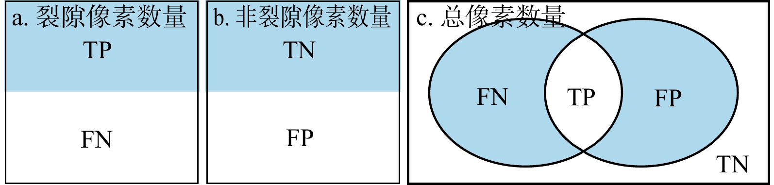
图 5 评价指标参数关系
TP为真正例,裂隙被正确分割为裂隙的像素数量;TN为真反例,非裂隙被正确分割为非裂隙的像素数量;FP为假正例,非裂隙被错误分割为裂隙的像素数量;FN为假反例,裂隙被错误分割为非裂隙的像素数量
Figure 5. Relationships among evaluation indicator parameters
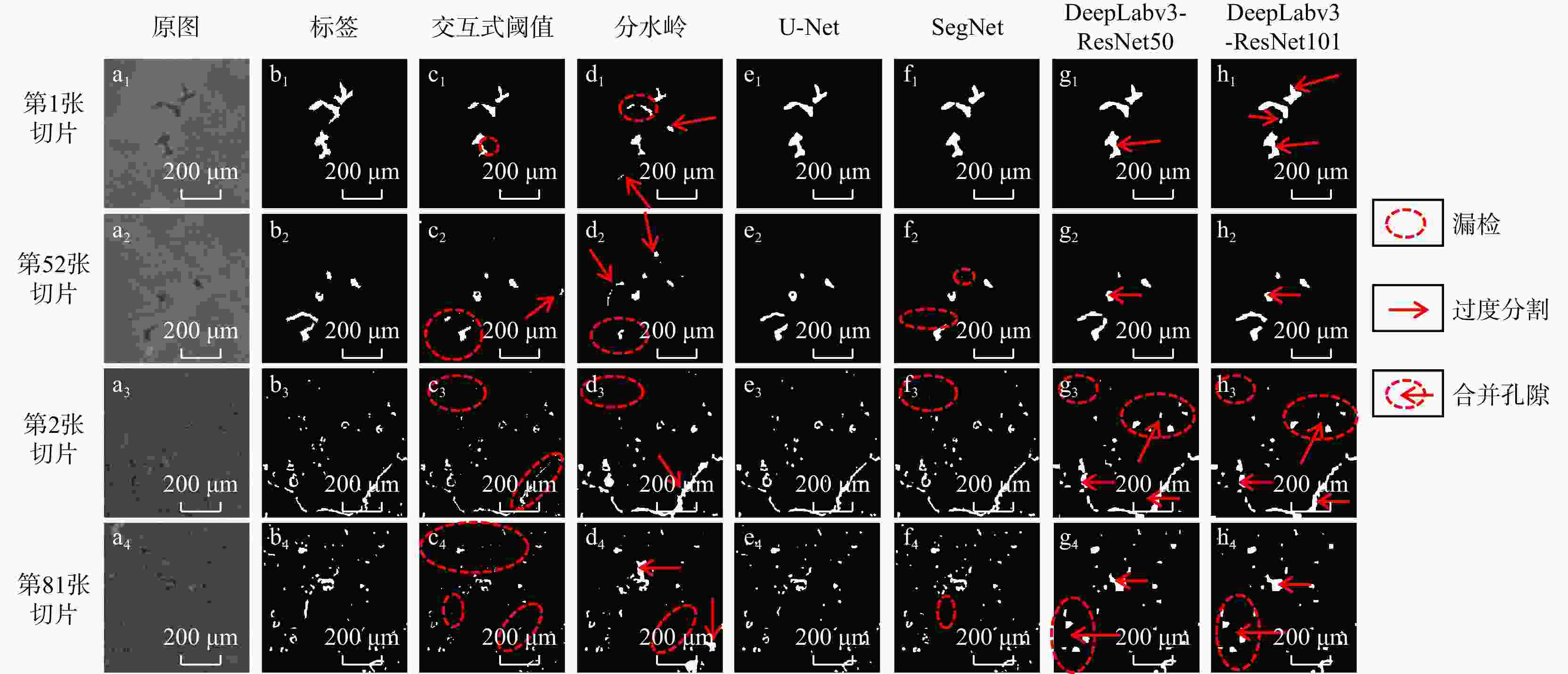
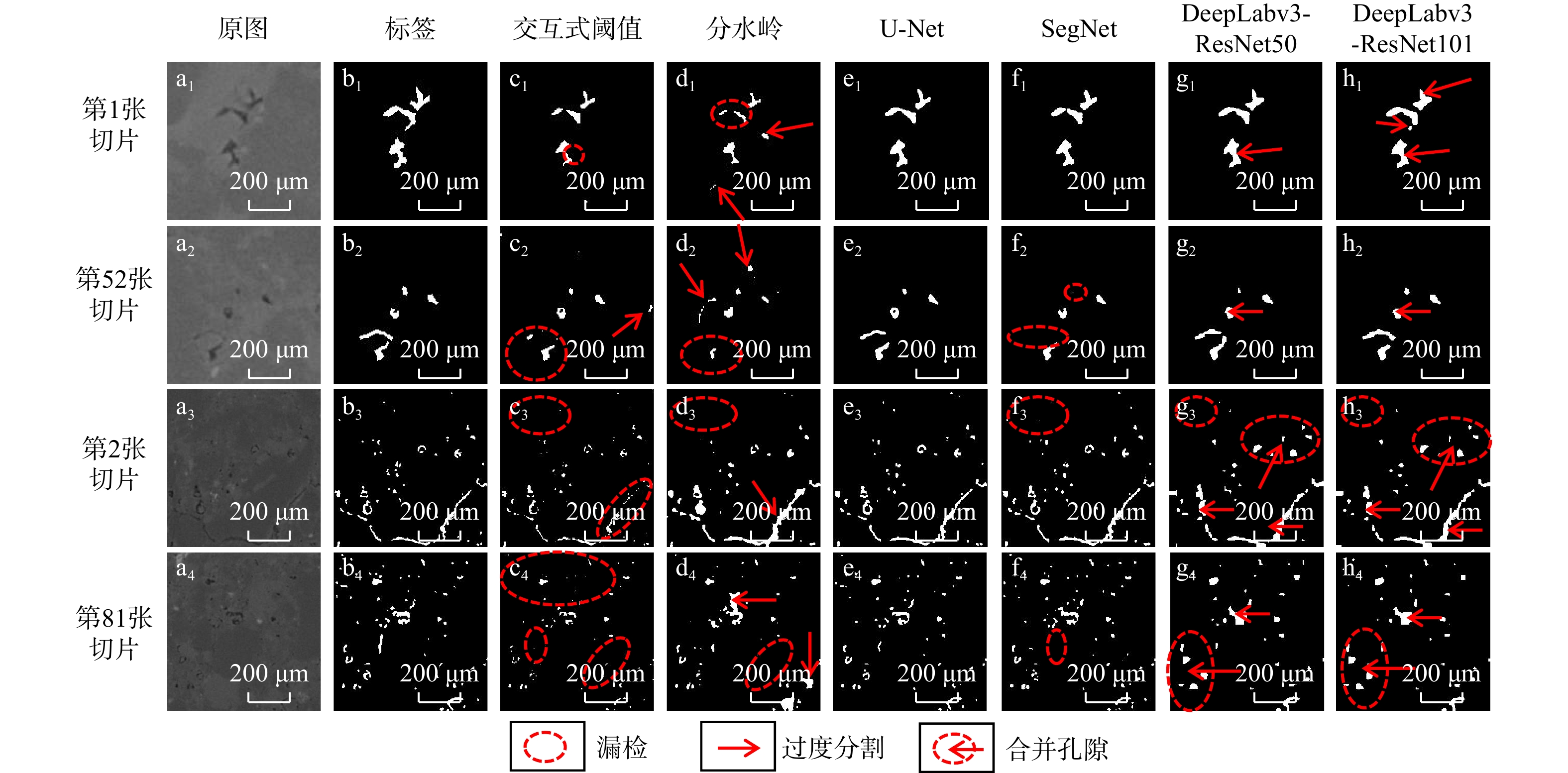
图 6 典型分割结果对比图(a1~h1,a2~h2为数据集一;a3~h3,a4~h4为数据集二)
Figure 6. Comparison of representative segmentation results
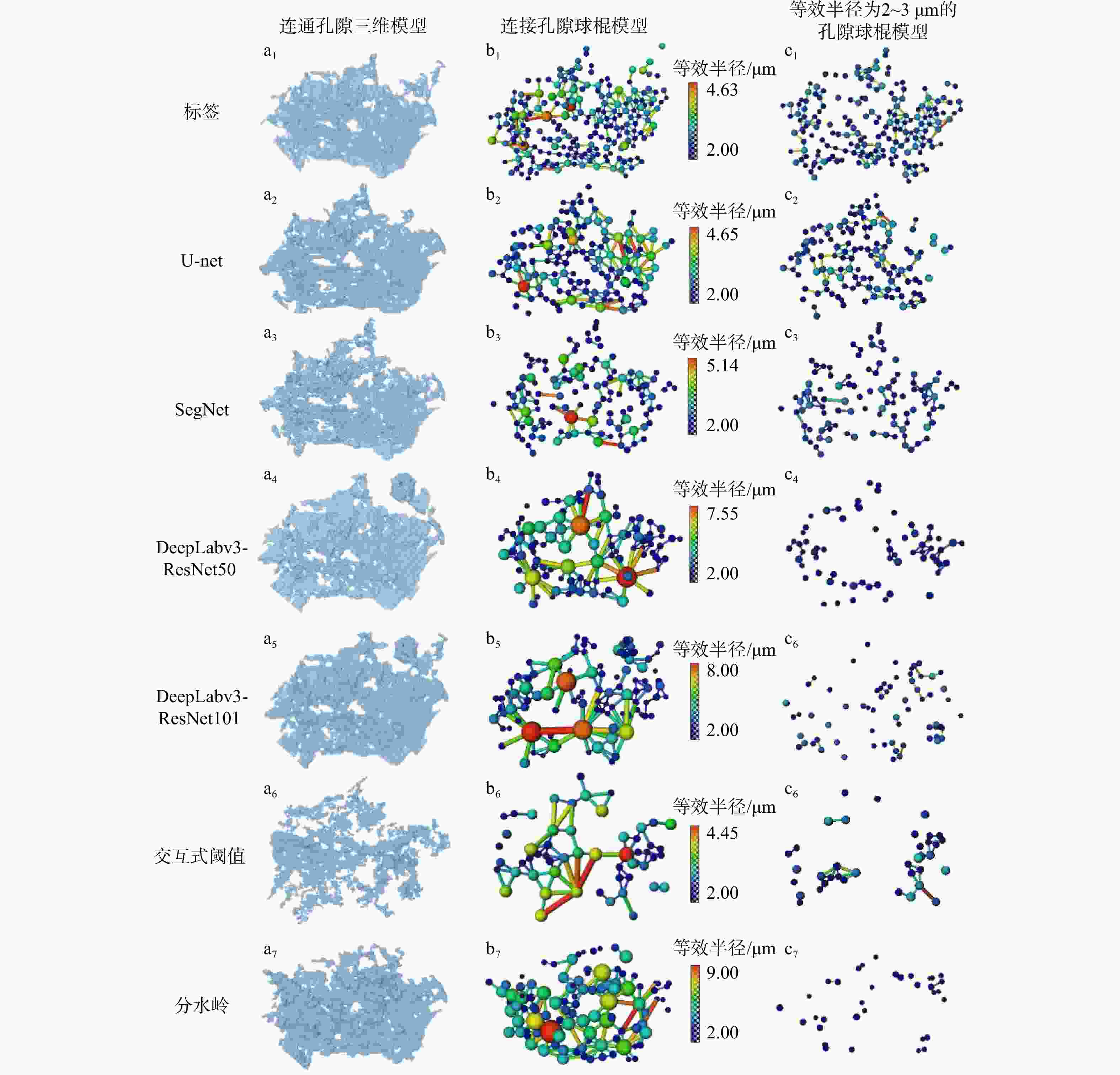
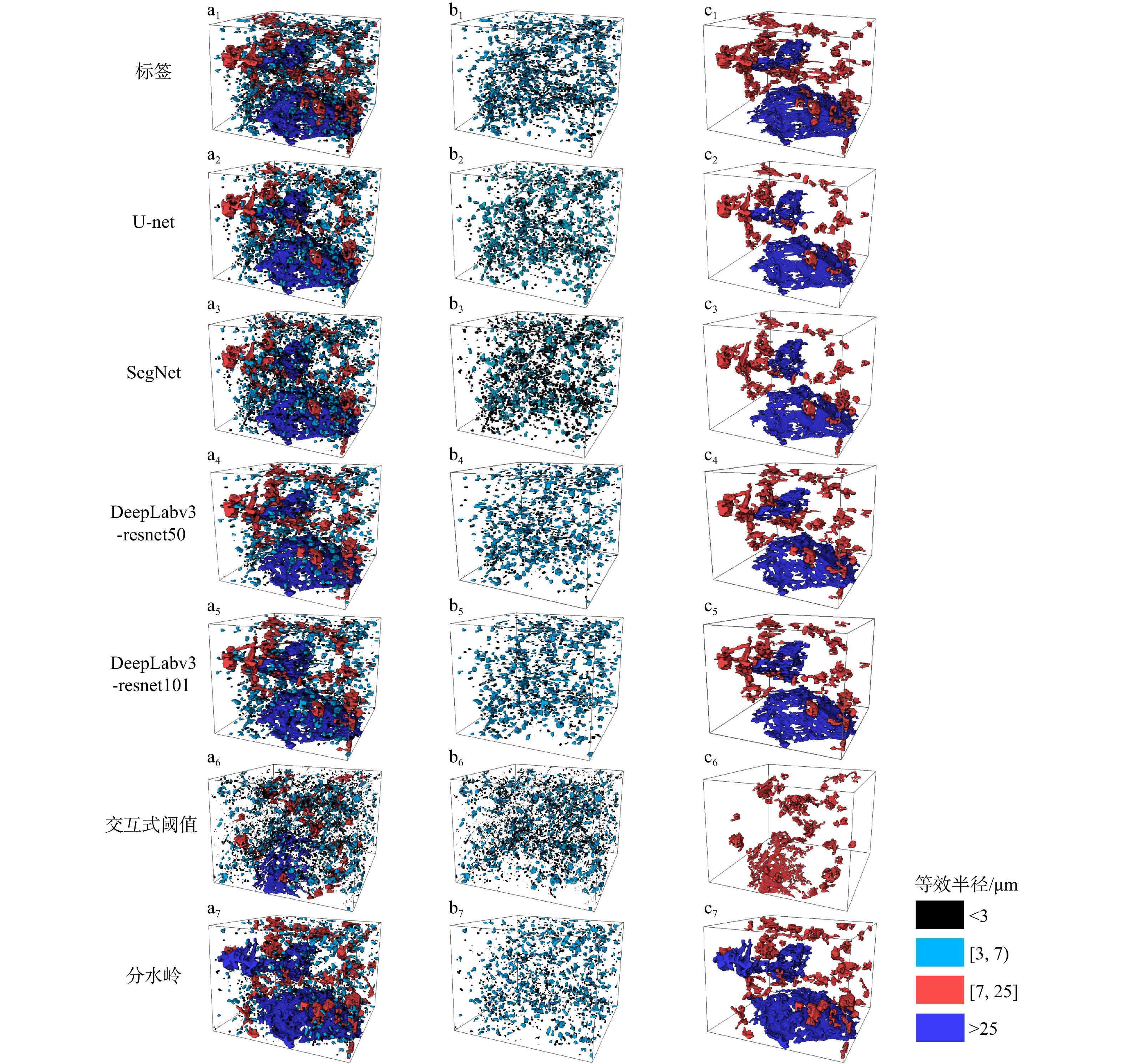
图 9 连通裂隙及其孔隙网络球棍模型
Figure 9. Connected fractures and corresponding ball-and-stick model of pore network
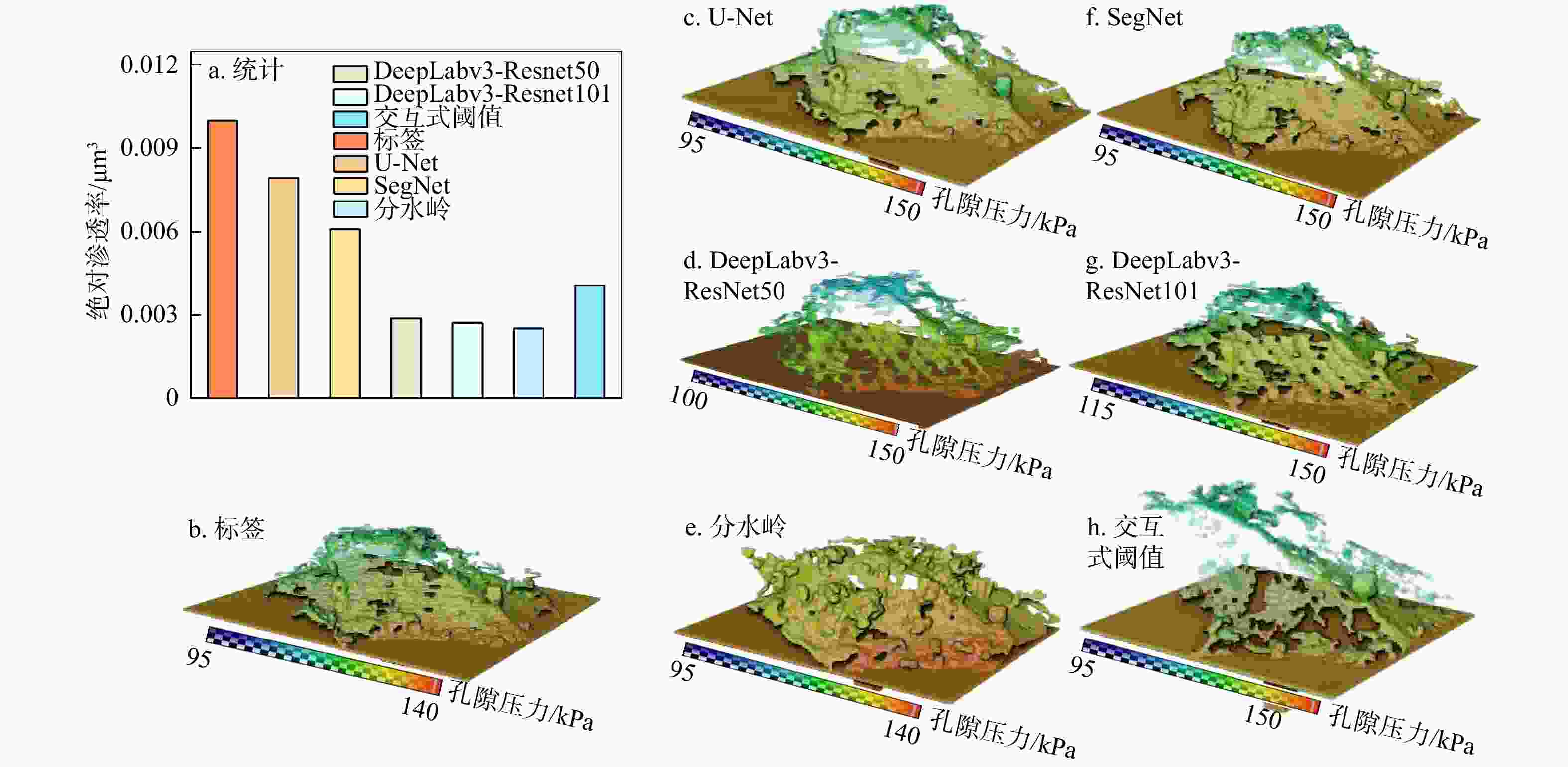
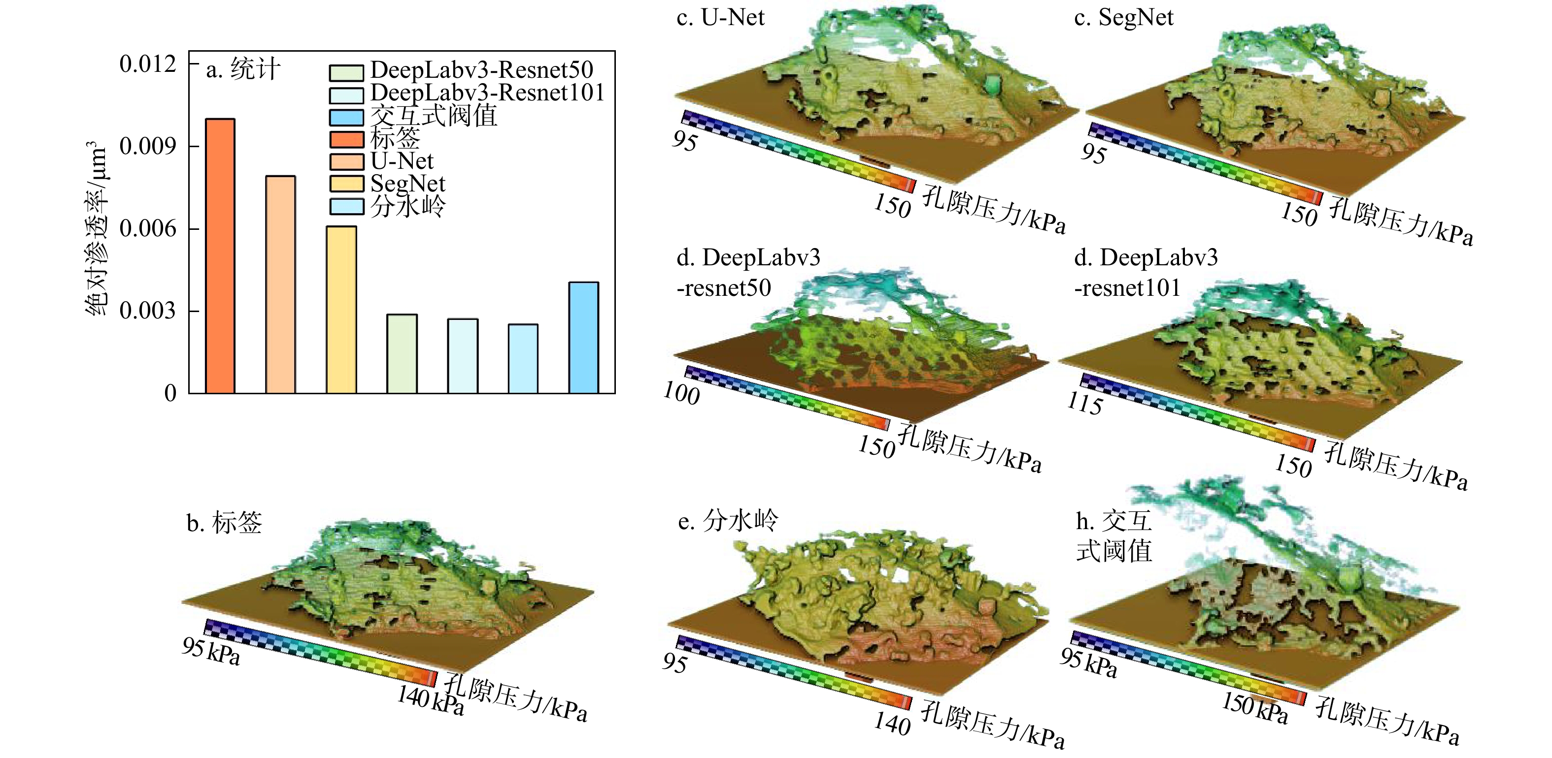
图 10 绝对渗透率模拟结果(a)及孔隙压力场分布(b~h)
Figure 10. Simulated absolute permeability results (a) and pore pressure field distribution (b-h)
表 1 主要矿物组成及其密度
Table 1. Major mineral composition and density
矿物名称 wB/% 密度/(g·cm−3) 矿物名称 wB/% 密度/(g·cm−3) 蒙脱石 36.60 2.00~3.00 石英 22.70 2.65 伊利石 2.79 方解石 16.80 2.71 高岭石 2.61~2.68 斜长石 15.30 5.54  下载: 导出CSV
表 2 各分割模型评价指标统计
Table 2. Statistics of evaluation indicators for each segmentation model
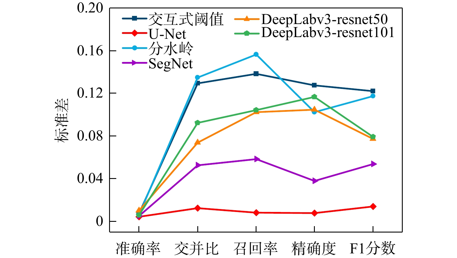
分割方法 评价指标/% 交并比IoU 准确率A 召回率R 精确率P F1分数F1 交互式阈值 40.74 98.27 42.96 89.08 57.70 分水岭 39.73 97.17 51.17 65.85 56.47 U-Net 59.44 99.03 70.84 79.98 74.17 SegNet 55.54 98.88 70.27 73.92 70.95 DeepLabv3-ResNet50 48.06 98.52 67.93 61.95 63.93 DeepLabv3-ResNet101 48.34 98.60 65.26 64.18 64.12
下载: 导出CSV
表 3 引入图像增强后各模型评价指标
Table 3. Evaluation indicators of different models with image enhancement
分割方法 评价指标/% 交并比IoU 准确率A 召回率R 精确率P F1分数F1 U-Net 53.68 98.18 61.99 69.96 65.13 SegNet 49.31 98.09 60.39 62.73 62.37 DeepLabv3-ResNet50 40.35 97.75 52.97 51.54 52.94 DeepLabv3-ResNet101 40.41 97.43 51.62 51.23 53.46
下载: 导出CSV
表 4 公开数据集下各模型性能评价
Table 4. Performance evaluation of different models on public dataset
分割方法 评价指标/% 交并比IoU 准确率A 召回率R 精确率P F1分数F1 U-Net 98.78 99.07 99.41 99.37 99.39 SegNet 99.18 98.61 99.10 99.06 99.08 DeepLabv3-resnet50 83.65 85.92 94.92 87.58 91.10 DeepLabv3-resnet101 83.60 85.90 94.68 87.72 91.06
下载: 导出CSV
表 5 孔隙网络模型(PNM)定量参数统计
Table 5. Quantitative parameter statistics of pore network model (PNM)
分割方法 连通孔
隙率/%孔 喉 数量 等效半径/μm 数量 吼道长度/μm 标签 0.67 440 2.07 1006 6.17 交互式阈值 0.21 100 2.12 174 7.81 分水岭 1.36 274 2.32 596 7.97 U-Net 0.61 387 2.03 630 6.32 SegNet 0.44 282 1.77 894 5.29 DeepLabv3-ResNet50 0.70 200 2.24 396 8.60 DeepLabv3-ResNet101 0.70 184 2.43 365 9.04
下载: 导出CSV
-
[1] 何云, 王瑞飞, 张占杨, 等. 鄂尔多斯盆地杭锦旗地区下石盒子组致密砂岩储层孔隙结构及可动流体特征[J]. 地质科技通报, 2023, 42(5): 94-102. doi: 10.19509/j.cnki.dzkq.2022.0134HE Y, WANG R F, ZHANG Z Y, et al. Pore structure and movable fluid characteristics of tight sandstone reservoirs in the Lower Shihezi Formation in the Hangjinqi area, Ordos Basin[J]. Bulletin of Geological Science and Technology, 2023, 42(5): 94-102. (in Chinese with English abstract) doi: 10.19509/j.cnki.dzkq.2022.0134 [2] 赵华伟, 廉培庆, 易杰, 等. 基于数字岩心技术的岩石孔渗特征研究: 以海外J油田孔隙型碳酸盐岩油藏为例[J]. 地质科技通报, 2023, 42(2): 347-355. doi: 10.19509/j.cnki.dzkq.tb20210522ZHAO H W, LIAN P Q, YI J, et al. A study of petrophysical properties based on digital core technology: A case study of a porous carbonate reservoir in the overseas J oilfield[J]. Bulletin of Geological Science and Technology, 2023, 42(2): 347-355. (in Chinese with English abstract) doi: 10.19509/j.cnki.dzkq.tb20210522 [3] 孟杰, 李长冬, 闫盛熠, 等. 基于μCT技术的白鹤滩库区致密砂岩孔−裂隙三维成像特征研究[J]. 地质科技通报, 2023, 42(1): 20-28. doi: 10.19509/j.cnki.dzkq.2022.0183MENG J, LI C D, YAN S Y, et al. 3D imaging characteristics of pore and fracture of tight sandstone in Baihetan reservoir area based on μCT technology[J]. Bulletin of Geological Science and Technology, 2023, 42(1): 20-28. (in Chinese with English abstract) doi: 10.19509/j.cnki.dzkq.2022.0183 [4] ZHAO G B, RUAN M D, TULU I B, et al. Reconstruct laminated shale cores/specimens for numerical modeling by physics-informed image processing[C]//Anon. 57th U. S. Rock Mechanics/Geomechanics Symposium. Atlanta, Georgia, USA: ARMA, 2023: ARMA-2023-0732. [5] 李元海, 徐晓华, 朱鸿鹄, 等. 基于计算机视觉的岩石裂隙识别表征与软件研制[J]. 岩土工程学报, 2024, 46(3): 459-469. doi: 10.11779/CJGE20221239LI Y H, XU X H, ZHU H H, et al. Identification and characterization of rock fractures based on computer vision and software development[J]. Chinese Journal of Geotechnical Engineering, 2024, 46(3): 459-469. (in Chinese with English abstract) doi: 10.11779/CJGE20221239 [6] 郑佳, 庄建琦, 孔嘉旭, 等. 基于CT扫描的黄土孔隙结构特征研究[J]. 地质科技通报, 2022, 41(6): 211-222. doi: 10.19509/j.cnki.dzkq.2022.0210ZHENG J, ZHUANG J Q, KONG J X, et al. Study of loess pore structure characteristics based on CT scanning[J]. Bulletin of Geological Science and Technology, 2022, 41(6): 211-222. (in Chinese with English abstract) doi: 10.19509/j.cnki.dzkq.2022.0210 [7] 朱红光, 谢和平, 易成, 等. 岩石材料微裂隙演化的CT识别[J]. 岩石力学与工程学报, 2011, 30(6): 1230-1238.ZHU H G, XIE H P, YI C, et al. CT identification of microcracks evolution for rock materials[J]. Chinese Journal of Rock Mechanics and Engineering, 2011, 30(6): 1230-1238. (in Chinese with English abstract) [8] JOHNS R A, STEUDE J S, CASTANIER L M, et al. Nondestructive measurements of fracture aperture in crystalline rock cores using X ray computed tomography[J]. Journal of Geophysical Research: Solid Earth, 1993, 98(B2): 1889-1900. doi: 10.1029/92JB02298 [9] WANG C, PEI W S, ZHANG M Y, et al. Multi-scale experimental investigations on the deterioration mechanism of sandstone under wetting-drying cycles[J]. Rock Mechanics and Rock Engineering, 2021, 54(1): 429-441. doi: 10.1007/s00603-020-02257-2 [10] 赵华伟, 宁正福, 段太忠, 等. 基于微米CT扫描成像实验及格子Boltzmann模拟方法的致密砂岩孔隙结构表征[J]. 东北石油大学学报, 2019, 43(5): 1-10. doi: 10.3969/j.issn.2095-4107.2019.05.001ZHAO H W, NING Z F, DUAN T Z, et al. Pore structure characterization of tight sandstones by X-ray computed tomography experiment combined with Lattice Boltzmann method[J]. Journal of Northeast Petroleum University, 2019, 43(5): 1-10. (in Chinese with English abstract) doi: 10.3969/j.issn.2095-4107.2019.05.001 [11] HE J M, LI T F, ZHENG B, et al. Effect of calcite veins on the mechanical behavior and fracture propagation of Carboniferous limestone[J]. Engineering Geology, 2022, 300: 106592. doi: 10.1016/j.enggeo.2022.106592 [12] RIVARD C, LAVOIE D, LEFEBVRE R, et al. An overview of Canadian shale gas production and environmental concerns[J]. International Journal of Coal Geology, 2014, 126: 64-76. doi: 10.1016/j.coal.2013.12.004 [13] ZHANG Y J. An overview of image and video segmentation in the last 40 years[M]//ZHANG Y J. Advances in Image and Video Segmentation. Hershey: IRM Press, 2006: 1-16. [14] 李晨安, 李承峰, 刘昌岭, 等. CT图像法计算Berea砂岩孔隙度[J]. 核电子学与探测技术, 2017, 37(5): 482-487.LI C A, LI C F, LIU C L, et al. A method to calculate the porosity of Berea sandstone based on CT digital images[J]. Nuclear Electronics & Detection Technology, 2017, 37(5): 482-487. (in Chinese with English abstract) [15] 陈超, 魏彪, 梁婷, 等. 一种基于工业CT技术的岩芯样品孔隙度测量分析方法[J]. 物探与化探, 2013, 37(3): 500-507.CHEN C, WEI B, LIANG T, et al. The application of industrial computation tomography (CT) to the analysis of core sample porosity[J]. Geophysical and Geochemical Exploration, 2013, 37(3): 500-507. (in Chinese with English abstract) [16] ALIZADEH S M, LATHAM S, MIDDLETON J, et al. An analysis of sleeve effects for petrophysical measurements using digital core analysis[C]//Anon. International Petroleum Technology Conference. Doha, Qatar: IPTC, 2015: D041S052R002. [17] AIT SKOURT B, EL HASSANI A, MAJDA A. Lung CT image segmentation using deep neural networks[J]. Procedia Computer Science, 2018, 127: 109-113. doi: 10.1016/j.procs.2018.01.104 [18] REINHARDT M, JACOB A, SADEGHNEJAD S, et al. Benchmarking conventional and machine learning segmentation techniques for digital rock physics analysis of fractured rocks[J]. Environmental Earth Sciences, 2022, 81(3): 71. doi: 10.1007/s12665-021-10133-7 [19] ALZUBAIDI F, MOSTAGHIMI P, SI G Y, et al. Automated rock quality designation using convolutional neural networks[J]. Rock Mechanics and Rock Engineering, 2022, 55(6): 3719-3734. doi: 10.1007/s00603-022-02805-y [20] NIU Y F, MOSTAGHIMI P, SHABANINEJAD M, et al. Digital rock segmentation for petrophysical analysis with reduced user bias using convolutional neural networks[J]. Water Resources Research, 2020, 56(2): e2019WR026597. doi: 10.1029/2019WR026597 [21] WU Y F, XIAO Z W, LI J, et al. ShaleSeg: Deep-learning dataset and models for practical fracture segmentation of large-scale shale CT images[J]. International Journal of Rock Mechanics and Mining Sciences, 2024, 180: 105820. doi: 10.1016/j.ijrmms.2024.105820 [22] LI B K, NIE X, CAI J C, et al. U-Net model for multi-component digital rock modeling of shales based on CT and QEMSCAN images[J]. Journal of Petroleum Science and Engineering, 2022, 216: 110734. doi: 10.1016/j.petrol.2022.110734 [23] SAFARI H, BALCOM B J, AFROUGH A. Characterization of pore and grain size distributions in porous geological samples: An image processing workflow[J]. Computers & Geosciences, 2021, 156: 104895. doi: 10.1016/j.cageo.2021.104895 [24] WANG H S, DALTON L, FAN M, et al. Deep-learning-based workflow for boundary and small target segmentation in digital rock images using U-Net++ and IK-EBM[J]. Journal of Petroleum Science and Engineering, 2022, 215: 110596. doi: 10.1016/j.petrol.2022.110596 [25] WANG F Y, ZAI Y. Image segmentation and flow prediction of digital rock with U-Net network[J]. Advances in Water Resources, 2023, 172: 104384. doi: 10.1016/j.advwatres.2023.104384 [26] ALQAHTANI N J, NIU Y F, WANG Y D, et al. Super-resolved segmentation of X-ray images of carbonate rocks using deep learning[J]. Transport in Porous Media, 2022, 143(2): 497-525. doi: 10.1007/s11242-022-01781-9 [27] RONNEBERGER O, FISCHER P, BROX T. U-Net: Convolutional networks for biomedical image segmentation[C]//Anon. Medical Image Computing and Computer-Assisted Intervention-MICCAI 2015. Cham: Springer, 2015: 234-241. [28] BADRINARAYANAN V, KENDALL A, CIPOLLA R. SegNet: A deep convolutional encoder-decoder architecture for image segmentation[J]. IEEE Transactions on Pattern Analysis and Machine Intelligence, 2017, 39(12): 2481-2495. doi: 10.1109/TPAMI.2016.2644615 [29] CHEN L C, PAPANDREOU G, KOKKINOS I, et al. Semantic image segmentation with deep convolutional nets and fully connected CRFs[C]//Anon. International Conference on Learning Representations. San Diego, USA: ICLR, 2015. [30] CHEN L C, PAPANDREOU G, KOKKINOS I, et al. DeepLab: Semantic image segmentation with deep convolutional nets, atrous convolution, and fully connected CRFs[J]. IEEE Transactions on Pattern Analysis and Machine Intelligence, 2018, 40(4): 834-848. doi: 10.1109/tpami.2017.2699184 [31] CHEN L C, ZHU Y K, PAPANDREOU G, et al. Encoder-decoder with atrous separable convolution for semantic image segmentation[C]//Anon. Computer Vision-ECCV 2018. Cham: Springer, 2018: 833-851. [32] FU J, LIU J, TIAN H J, et al. Dual attention network for scene segmentation[C]//Anon. 2019 IEEE/CVF Conference on Computer Vision and Pattern Recognition (CVPR). Long Beach, CA, USA: IEEE, 2020: 3141-3149. [33] PAL D, REDDY P B, ROY S. Attention UW-Net: A fully connected model for automatic segmentation and annotation of chest X-ray[J]. Computers in Biology and Medicine, 2022, 150: 106083. doi: 10.1016/j.compbiomed.2022.106083 [34] WU Y, JIANG J Y, HUANG Z M, et al. FPANet: Feature pyramid aggregation network for real-time semantic segmentation[J]. Applied Intelligence, 2022, 52(3): 3319-3336. doi: 10.1007/s10489-021-02603-z [35] BAZAIKIN Y, GUREVICH B, IGLAUER S, et al. Effect of CT image size and resolution on the accuracy of rock property estimates[J]. Journal of Geophysical Research: Solid Earth, 2017, 122(5): 3635-3647. doi: 10.1002/2016JB013575 [36] LAI P, MOULTON K, KREVOR S. Pore-scale heterogeneity in the mineral distribution and reactive surface area of porous rocks[J]. Chemical Geology, 2015, 411: 260-273. doi: 10.1016/j.chemgeo.2015.07.010 [37] WANG Y Z, RAHMAN S S, ARNS C H. Super resolution reconstruction of μ-CT image of rock sample using neighbour embedding algorithm[J]. Physica A: Statistical Mechanics and its Applications, 2018, 493: 177-188. doi: 10.1016/j.physa.2017.10.022 [38] SINGH A, REGENAUER-LIEB K, WALSH S D C, et al. On representative elementary volumes of grayscale micro-CT images of porous media[J]. Geophysical Research Letters, 2020, 47(15): e2020GL088594. doi: 10.1029/2020GL088594 [39] LECUN Y, BOTTOU L, BENGIO Y, et al. Gradient-based learning applied to document recognition[J]. Proceedings of the IEEE, 1998, 86(11): 2278-2324. doi: 10.1109/5.726791 [40] HE K M, ZHANG X Y, REN S Q, et al. Deep residual learning for image recognition[C]//Anon. 2016 IEEE Conference on Computer Vision and Pattern Recognition (CVPR). Las Vegas, NV, USA: IEEE, 2016: 770-778. [41] RABBANI A, JAMSHIDI S, SALEHI S. An automated simple algorithm for realistic pore network extraction from micro-tomography images[J]. Journal of Petroleum Science and Engineering, 2014, 123: 164-171. doi: 10.1016/j.petrol.2014.08.020 [42] MOSTAGHIMI P, BLUNT M J, BIJELJIC B. Computations of absolute permeability on micro-CT images[J]. Mathematical Geosciences, 2013, 45(1): 103-125. doi: 10.1007/s11004-012-9431-4 [43] SONG R, WANG Y, LIU J, et al. Comparative analysis on pore‐scale permeability prediction on micro-CT images of rock using numerical and empirical approaches[J]. Energy Science & Engineering, 2019, 7(6): 2842-2854. doi: 10.1002/ese3.465 [44] WANG G, QIN X J, HAN D Y, et al. Study on seepage and deformation characteristics of coal microstructure by 3D reconstruction of CT images at high temperatures[J]. International Journal of Mining Science and Technology, 2021, 31(2): 175-185. doi: 10.1016/j.ijmst.2020.11.003 [45] DONG J X, DAI P X, LIU W L, et al. Characterizing disintegrated dolomite pore structure and seepage: CT scanning and numerical approach[J]. Journal of Rock Mechanics and Geotechnical Engineering, 2025, 17(7): 4386-4399. doi: 10.1016/j.jrmge.2024.11.013 [46] RAMANDI H L, MOSTAGHIMI P, ARMSTRONG R T. Digital rock analysis for accurate prediction of fractured media permeability[J]. Journal of Hydrology, 2017, 554: 817-826. doi: 10.1016/j.jhydrol.2016.08.029 [47] ZHAO X X, WANG X. Unsupervised domain adaptation based fracture segmentation method for core CT images[J]. Expert Systems with Applications, 2025, 264: 125857. doi: 10.1016/j.eswa.2024.125857 -

 下载:




 点击查看大图
计量
- 文章访问数: 597
- PDF下载量: 60
- 被引次数: 0